Paget’s Disease of the Breast
By: N. Mai Bui, BA and Daniel Bradley, MD
Paget’s disease of the breast is a form of breast cancer involving the skin of the nipple and areola. This is a rare condition and has been estimated to represent 1-4% of all breast cancers. Paget’s disease of the breast most commonly occurs in postmenopausal women, with a mean age of 57. Paget’s disease of the breast is associated with an underlying cancer in other areas of the breast in over 90% of cases.
Most patients with Paget’s disease of the breast present with a red, scaly rash on the skin of the nipple and/or areola. The affected area is often painful and may also be associated with an itching or burning sensation. These symptoms may be similar to those found in benign skin conditions, such as eczema or psoriasis. Bloody or serous nipple discharge and retraction of the nipple can also be seen. A palpable mass in the subareolar area may also be appreciated on physical exam.
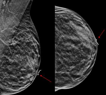
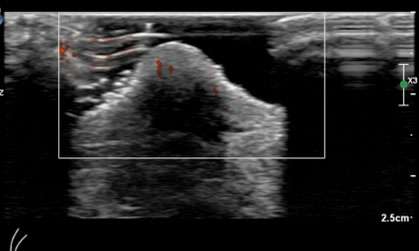
As Paget’s disease of the breast may be multicentric, it is essential to evaluate the entirety of the breast outside of the nipple/areola region to assess for additional sites of tumor. Mammogram findings of Paget’s disease of the breast include thickening of the skin at the nipple and areola, nipple retraction/inversion, microcalcifications, masses, or architectural distortion. However, a mammographic correlate may not always be seen. Ultrasound is often helpful for further evaluation when the mammogram is negative or when patients complain of a palpable lump. Ultrasound findings may include skin thickening of the nipple, heterogenous hypoechoic areas, areas of increased vascularity, discrete masses, and dilated ducts.
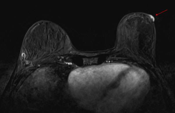
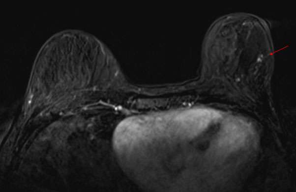
Breast MRI is highly sensitive for the detection of breast cancer and may be used in patients with Paget’s disease of the breast to establish the extent of disease. MRI may show thickening of the nipple/areola, asymmetric abnormal enhancement of the nipple/areola, enhancing masses, and linear/segmental non-mass enhancement.
References:
- Karakas C. "Paget's Disease of the Breast." J Carcinog. 2011;10:31. doi: 10.4103/1477-3163.90676. Epub 2011 Dec 8. PMID: 22279416; PMCID: PMC3263015.
- Sripathi S, Ayachit A, Kadavigere R, Kumar S, Eleti A, Sraj A. "Spectrum of Imaging Findings in Paget's Disease of the Breast-A Pictorial Review." Insights Imaging. 2015 Aug;6(4):419-29. doi: 10.1007/s13244-015-0415-z. Epub 2015 Jul 5. PMID: 26142549; PMCID: PMC4519816.