Prostate Imaging
Small Volume Low Grade Disease
Find your care
Our radiologists lead the way in prostate imaging. We offer the newest techniques to better detect and stage prostate cancer. Call 310-481-7545 to find out more about prostate imaging and treatment options.
History
- 60 year-old
- PSA 3.6
- 5 of 6 right biopsies GS3+3
Imaging
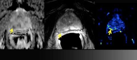
LEFT: Apparent diffusion coefficient (ADC) map: moderately restricted diffusion
CENTER: T2 Weighted imaging: ill-defined low signal (yellow arrow) does not abut the prostate capsule
RIGHT: Perfusion map: focally increased perfusion
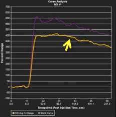
LEFT: Perfusion time-intensity curve: type III (washout) curve is highly suspicious
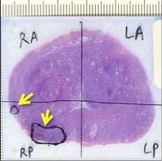
RIGHT: Whole-mount pathology: two low-grade (Gleason Score 3+3=6) tumors do not involve capsule
Advantage: UCLA Prostate MRI
- Although less sensitive for low-grade disease, MRI can help with determining extent and determination whether surveillance or definitive treatment is warranted.
- Diffusion weighted imaging (such as Siemens REVEAL) with multiple b-values allows for high contrast-to-background on ADC map and computation of high b-value images