Prostate Imaging
MR-Guided Targeted Biopsy
Find your care
Our radiologists lead the way in prostate imaging. We offer the newest techniques to better detect and stage prostate cancer. Call 310-481-7545 to find out more about prostate imaging and treatment options.
History
- 63 y/o, PSA 8.8 → 13.2 over 5 years
- All systematic biopsies negative
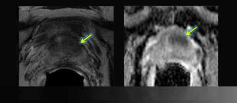
- Hypointense left anterior lesion with restricted diffusion is moderately suspicious, not in biopsy zone
Imaging

LEFT: Axial T2-weighted image: asymmetric anterior low signal
RIGHT: Apparent diffusion coefficient (ADC) map: focal restricted diffusion


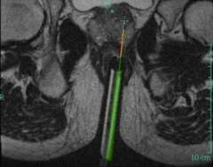
LEFT: Oblique Axial T2-weighted image from in-bore MR-guided targeting
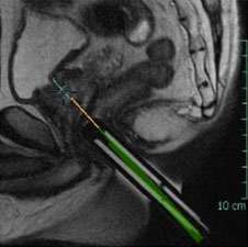
RIGHT: Corresponding sagittal T2-weighted image from in-bore MR-guided targeting
Results
- Gleason grade 3+3=6/10
- All biopsies over 5 mm and over 50% of core
Advantage: UCLA Prostate MRI
Suspicious findings on diagnostic multiparametric prostate MRI can be targeted for direct in-bore MRI-guided biopsy or MRI-ultrasound fusion targeted biopsy