Prostate Imaging
Extracapsular Extension and Seminal Vesicle Invasion
Find your care
Our radiologists lead the way in prostate imaging. We offer the newest techniques to better detect and stage prostate cancer. Call 310-481-7545 to find out more about prostate imaging and treatment options.
History
- 59 year-old male.
- PSA level: 10
- Clinical staging: T1C
- Blind systematic biopsy results: Gleason: 3+4=7 in the Left prostate
Imaging
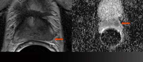
LEFT: Axial T2-weighted image: low signal bulges the capsule (arrow)
RIGHT: Gray-scale apparent diffusion coefficient (ADC) map: focal restricted diffusion (arrow)
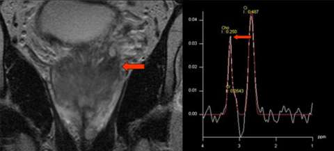
LEFT: Coronal T2-weighted image: abnormal signal extends to seminal vesicles (arrow)
RIGHT: Sample spectrum from spectroscopic imaging: elevated choline (Cho) relative to citrate (Ci)
Results
- Gleason Score 3+4 prostate cancer
- Extra-capsular extension,
- Left seminal vesicle invasion.
- TNM staging : T3b
Advantage: UCLA Prostate MRI
For patients at intermediate risk of T3 disease, MRI can stratify risk.