Diagnosis and Staging
Find your care
A team of experts collaborates to provide advanced mesothelioma care. Call 310-267-4612 to learn more about mesothelioma treatment at UCLA Health. To reach our nurse practitioner, call 310-818-1304
Diagnosis and staging of malignant pleural mesothelioma
Our expert team uses state-of-the-art technology and precise procedures to diagnose mesothelioma and see how far it has spread. With this information, we create a personalized treatment plan tailored to each patient.
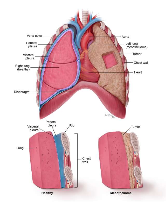
Healthy lung compared to mesothelioma
Diagnosis
Diagnosing malignant pleural mesothelioma often begins with imaging tests followed by procedures to collect tissue samples. These tests help confirm the presence of cancer and identify where it is located.
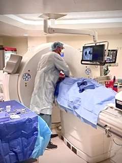
CT Scan
A CT scan is commonly used to help diagnose mesothelioma by providing detailed images of the chest or abdomen.
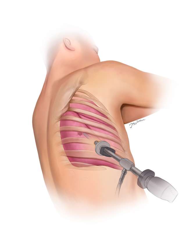
CT guided core needle biopsy
This is a procedure when a needle is inserted into the tumor to sample it, under guidance of a CT scan. It is often indicated for larger tumors. More information.
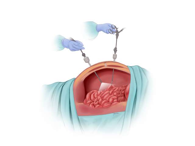
Thoracoscopy with biopsy
This minimally invasive surgical procedure uses a small camera inserted through a tiny incision in the chest and has high accuracy for diagnosing mesothelioma. Pleurodesis is often performed during the same procedure to treat pleural effusion, which is a buildup of fluid in the chest cavity.
Staging
Staging helps determine how far mesothelioma has spread and plays a key role in choosing the right treatment. The most common system used is the TNM staging system, developed by the American Joint Committee on Cancer (AJCC):
- T (Tumor) describes the size and location of the main tumor
- N (Node) shows whether nearby lymph nodes are involved
- M (Metastasis) indicates if the cancer has spread to other parts of the body
The TNM system is not a test itself, but a way to categorize the cancer using results from various diagnostic procedures. The following tests are commonly used to help determine the stage of mesothelioma:
MRI
An MRI is sometimes used to help stage mesothelioma by showing detailed images of soft tissues. It can help detect if the cancer has spread to areas like the chest wall, diaphragm, or muscles. This information is useful for planning surgery or other treatments
Integrated PET-CT scan
This test combines two powerful scans into one: a CT scan and a PET scan. The CT shows detailed pictures of the body, and the PET scan shows areas where the cancer is active. Together, they help doctors see how far the cancer has spread. More information.
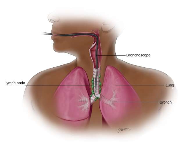
Endobronchial ultrasound (EBUS)
EBUS is a less invasive way to examine and biopsy lymph nodes in the chest. A special camera with ultrasound is passed through the airways to reach the lymph nodes and take a sample.
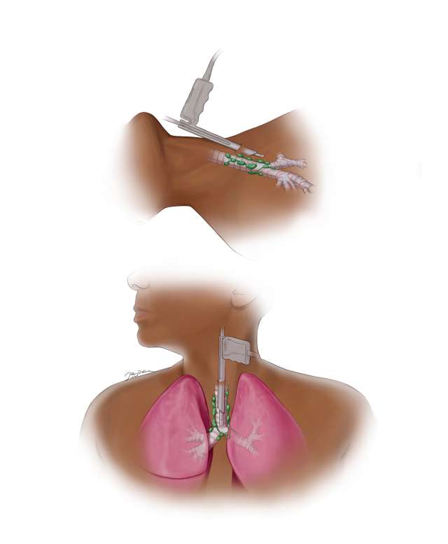
Cervical mediastinoscopy
This is a short procedure where doctors make a small cut in the lower neck to look at and biopsy the lymph nodes in the middle of the chest. This helps check whether the cancer has spread beyond the lungs to the lymph nodes.
Diagnostic laparoscopy
Sometimes mesothelioma can spread from the chest to the belly. In this procedure, a small camera is placed through a tiny cut in the abdomen to check for signs of cancer in the abdominal area.