Our Research
Bone Biology
Our Goal is to develop more sensitive in vitro and in vivo biological tests. The laboratory is currently developing a new animal model to evaluate osteolyitc potential of UHMWPE particles.
Wearable Technologies
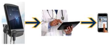
Joint and Spine Monitoring Devices
- Based on AE (ultrasound) Technology
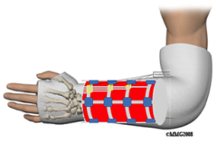
Smart Cast
- Based on continuous pressure and O2 perfusion monitoring
- Fracture healing stimulation based on ultrasound
Anti-infection Enhancing Devices
- Based on external stimulation via ultrasound or micro-currents
- Smart Nanocoating Activation
- Drug Delivery Modulation

Gait Analysis
- Based on wireless IMU and EMG sensors
Biomaterials & Biomechanics
Biomaterials, Biomechanics and Tribology
Scope
The goal of research in the Tribology, Biomechanics and Biomaterials Laboratory is to develop more durable bearing surfaces to extend their useful lifespan in patients and develop new materials and surface treatments for orthopaedic implants. The laboratory is also fully equipped to test and evaluate new third parties implants and materials.
Research
Studies based in the Tribology Laboratory include:
- Evaluation of existing or prototype materials/devices for orthopaedic implants.
- Development of new, improved materials & devices.
- Evaluation of novel and explanted implants.
Nanoparticles
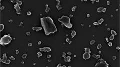
Accurate Nanoparticle Characterization
In the past accurate analysis of wear particles has been limited by problems associated with particle isolation, distribution, and display. For example, an inefficient and/or incomplete digestion of the proteinaceous content can lead to agglomeration or loss of particles. Furthermore, separation of particles from digested solution through filtration or embedding into resins can be an additional source of agglomeration and loss.
Moreover particles of small dimension (few nanometers) can easily bind to proteins, stick to the wall of tubes or flushed away through the pores of filters. Agglomeration, loss, unwanted residues affect the quality and clarity of particle imaging with obvious repercussions on the reliability and reproducibility of the morphological analysis.
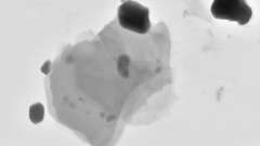
Failure Analysis
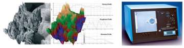
Failure Analysis of Implants and Components
We study and determine the mode of failure of explanted implants using a range of specifically designed tests. The large database of information from previously analyzed implant revisions allow us for a more rapid and precise identification of the reasons for any subsequent revisions.
What we offer
- Macro and microscopic analysis of implant surfaces;
- Image analysis of in/ongrowth surfaces (LM, SEM);
- Fractography of structural failures (SEM);
- X-Ray investigative techniques (XPS, EDS);
- Wear assessment of bearing interfaces (CMM, Profilometer combined with a 3-D image software, Wear Scar Mapping);
- Corrosion Assessment (SEM, EDS, XPS).
Core Facilities
-
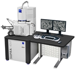
Field Emission Scanning Electron Microscopy: Zeiss Supra 40 VP (Peltier Stage, SE, BSD, In-Lens, and STEM detectors; EDS, Alicona MeX 3D metrology software)
- Ion Beam Sputter Coater: IBSE (Ir, C, Pt-Pd, Au)
- Optical Microscopy: Zeiss, Leica
- Microtomes
Funding

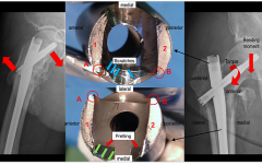
1R43AR067048-01A1 – “Rapid Detection of Common Failure Modes for Knee Prostheses”
1R21AR069287-01 “Real-time Monitoring of Knee Injuries”
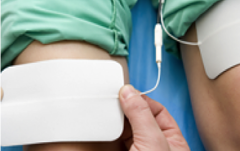
Effect of PEMF on Osteogenesis.