Small Animal Imaging
Overview
The Small Animal Imaging Shared Resource (SAISR) provides state-of-the-art in vivo imaging technologies and related diagnostic and radio-therapeutic services to JCCC and UCLA faculty, staff and students. The SAISR is a world-class, first-of-its-kind small animal imaging technology development and shared resources facility, providing JCCC investigators with technical imaging expertise and facilitating the design, execution and analysis of in vivo small animal imaging studies.
Our aims are:
- To provide expertise and support state-of-the-art small animal imaging technologies
- To provide expertise and support radiochemistry and radiolabeling production services
- To provide JCCC investigators with training in radiochemistry, preclinical imaging, and image analysis and to support the design and execution of imaging studies
The SAISR offers full-service whole body microPET, microCT, MRI, and optical imaging and complementary in vitro/ex vivo assays to measure tissue function and structure. Companion PET tracer radiochemistry and radiolabeling services are available in-house through the adjacent radiochemistry and cyclotron facility. Study design and development, training in imaging techniques, and staff assistance are offered regularly to all users. Please contact the SAISR at [email protected] to get started.
Leadership


Mailing Address
Preclinical Imaging Technology Center
Crump Institute for Molecular Imaging
California NanoSystems Institute (CNSI)
2nd floor, Room 2112
Los Angeles, CA 90095-1770
(in the Court of Sciences)
Imaging Center
Phone: 310-794-2224
Email: [email protected]
Arion Chatziioannou: 310-825-7877
Shili Xu: 310-825-7137
Websites
Go to Preclinical Imaging Technology Center website
Go to Crump Institute for Molecular Imaging website

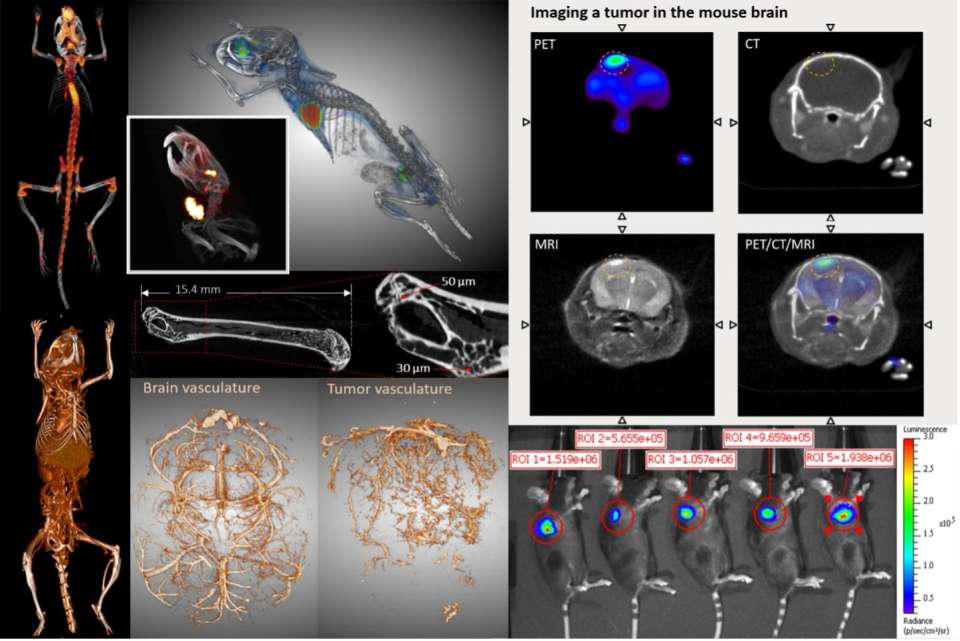
In Vivo Imaging Services
Nuclear imaging
- Positron emission tomography (microPET)
- Static and dynamic whole body functional imaging
- Extensive panel of radiolabeled small molecule PET tracers (readily available)
- Peptide/protein radiolabeling service and workspace
- Single Photon Emission Computed Tomography (SPECT)
- Whole body imaging of targeted radiopharmaceuticals
- Simultaneous multi-isotope imaging
- Computed tomography (microCT)
- Whole body anatomical imaging
- Quick scan time for in vivo imaging (1-2min)
- Down to 20 µm (specimen imaging) and 100 µm (in vivo imaging) resolution
- Contrast enhancement of soft tissues such as lymph nodes, spleen, liver, heart, gastrointestinal, major blood vessels
- Cardiac gating and respiratory gating are available
Optical imaging
- Bioluminescence, Fluorescence and Cerenkov imaging
- Whole body, multi-animal imaging
- Cell localization, target trafficking, longitudinal studies
- BLI: D-luciferin substrate provided
- FLI: multiple excitation/emission filters including near-infrared
MRI imaging
- Aspect 1T preclinical MRI system (50 micron resolution) with rodent head coil and body coil
- ECG-gated cardiac MRI available
Complementary services
- Routine productions of established PET tracers
- Novel PET tracer consultation and development
- Biologics radiolabeling
- Toxicology of new PET tracers for IND submission (through UCLA DLAM)
- Imaging-based pharmacokinetics and pharmacodynamics studies
- Radiotherapeutic labeling (e.g. Lu-177, Y-90) and efficacy studies
- PET tracer whole body biodistribution and dosimetry
- PET tracer metabolite analysis
- In vivo and ex vivo biodistribution studies of imaging agents
- In vitro binding and uptake assays (e.g. radiolabeled PET tracers; fluorescent biomarkers)
- In vitro and ex vivo (tissue or whole body) autoradiography
- Quantitative image analysis and training
- Image data processing and 2D slice or 3D volume renderings
- DICOM formatting and archiving
- 3D printing of CT-scanned objects
- Rodent tail vein i.v. injection of experimental agents and cells
Equipment
In vivo imaging
- GNEXT combined microPET/CT scanner (Sofie Biosciences)
- NanoScan SPECT/CT scanner (Mediso)
- In-house quick microCT scanner (CrumpCAT)
- M2 1T MRI (Aspect)
- IVIS Lumina II bioluminescent and fluorescent optical imaging system
Sample imaging
- High resolution (18 micron) CT scan of fixed tissues or samples.
Radiochemistry and Radiolabeling
- Siemens RDS-111 cyclotron
- multiple Sofie Biosciences ELIXYS FLEX/CHEM automated radiosynthesizers
- Raytest Ginastar radio-TLC reader
- multiple Knauer Smartline HPLC systems with Bioscan gamma detectors
- multiple Capintec CRC-25 PET dose calibrators
- Agilent gas chromatograph with mass spec detector
- Iodination chamber
- Cerenkov imaging system
Complementary instruments
- Wizard 3" automatic gamma counter (Perkin Elmer)
- 300SL liquid scintillation counter (HiDex)
- Leica CM3600 XP cryomacrotome – whole body sectioning
- Leica CM3050 cryomicrotome – tissue sectioning
- FUJIFILM BAS-500 phosphor imaging system
- Multi-nozzle FDM 3D printers and SLA 3D printers
- Agilent 1100 HPLC with UV/Vis detector, Bioscan Flow-Count radiation detector and fraction collector
- Cell culture facility: biosafety cabinet, incubator, microscope, centrifuge
- Adams MHCT II whole blood micro-hematocrit centrifuge – blood separation
- Microfuge 22R centrifuge (Beckman Coulter)
- Isoflurane vaporizers, induction chambers, nose cones, and animal heating stations
- Multiple BSL2-level biosafety cabinets and chemical fume hoods
- Adjacent full-service DLAM vivarium and procedure room for mice and rats (through DLAM)
Operations
- New investigators can contact the SAISR at [email protected] to discuss potential studies, experimental design, data analysis, and for questions or advice
- After training, JCCC researchers can readily use the SAISR through secure keycard access around the clock
- Experiments are generally performed by members of the investigator’s lab after SAISR training, with expertise and assistance from the SAISR faculty and staff
- Animal housing facility: Adjacent to the Imaging Center is a barrier facility for housing animals before and after imaging with biosafety cabinets for procedures
- The Imaging Center is fully certified by UCLA’s Office of Animal Research Oversight (OARO) and Environmental Health and Safety (EH&S) for animal welfare and radiation, laboratory, biological and chemical safety procedures
Pricing
| Service | JCCC Rate | Per Unit |
|---|---|---|
| microPET/CT imaging | $322.91 | Hour |
| SPECT/CT imaging | $239.87 | Hour |
| microCT imaging | $148.55 | Hour |
| MRI | $176.32 | Hour |
| Bioluminescence (includes D-luciferin) or fluorescence imaging | $108.14 | Hour |
| Usage of gamma counter, liquid scintillation counter, cryomacrotome, cryomicrotome, cryomacrotome, cryomicrotome, Fuji BAS phosphor imager, procedure workspace, etc. | $44 | Hour |
| Imaging staff assistance | $115.02 | Hour |
| Imaging data analysis | $164.41 | Hour |
| Tain vein i.v. injedction | $29.22 | Injection |
| 3D printing | $8.89 | Hour |
List of Available PET Tracers
| PET Probe | Type | Biological Application |
|---|---|---|
| [18F]FDG | Glucose | Glycolysis (oncology, neurology, etc.) |
| [18F]NaF | Hydroxyapatite | bone scan; cardiac calcification; oncology (e.g. prostate cancer bone metastasis) |
| [18F]FLT | Thymidine (nucleoside analog) | Proliferation; nucleoside metabolism (e.g. oncology, immunology) |
| [18F]FAC (and analogs) | Deoxycytidine (nucleoside analog) | Immunology (e.g. adaptive immune response; immunotherapies) oncology (e.g. treatment stratification of nucleoside analog prodrugs) |
| [18F]clofarabine | Purine (nucleoside analog | Immunology; oncology |
| [18F]AraG | Guanosine (nucleoside analog) | Immunology; oncology |
| [18F]FHBG, FEAU, L-FMAU, FIAU (and analogs) | Guanosine (nucieoside analog) | Immunology; oncology |
| [18F]FDHT | Testosterone | Oncology (prostate cancer) |
| [18F]FDOPA | Amino acid (phenylalanine) | Neuroscience (dopaminergic) oncology: brain tumors |
| [18F]fallypride | Dopamine receptor antagonist | Neuroscience (dopaminergic) |
| [18F]FDDNP | Ammonia | Routinely for clinical imaging: neurology (e.g., Alzheimer's disease) |
| [11C]acetate | Acetate | Oncology (prostate cancer) fatty acid synthesis |
| ImmunoPET (various radioisotopes) | Full antibodies and antibody fragments | Oncology, immunology, cell surface targets |
| [68Ga]DOTA-TOC | Somatostatin analogs | Oncology (e.g., neuroendocrine tumors) |
| [68Ga]DOTA-NOC | Somatostatin analogs | Oncology (e.g., neuroendocrine tumors) |
| [11C]choline | Choline | Oncology (prostate cancer) |
| [11C]L-glutamine | Glutamine | Oncology; glutamine metabolism |
| [18F]fluorobenzyl triphenylphosphonium ([18F]FBnTP) | Mitohondrial membrane potential | Oncology |
| [18F]florbetaben | Amyloid | Neuroscience |
Protein/Peptide Labeling
| Name | Group | Labeling |
|---|---|---|
| [18F]SFB | Prosthetic group | Peptide/protein labeling |
| [18F]FBAM | Prosthetic group | Peptide/protein labeling |
| [18F]FBEM | Prosthetic group | Peptide/protein labeling |
| [18F]fluorobenzaldehyde | Prosthetic group | Peptide/protein labeling |
| [18F]fluoroethyl-PEG3-azide | Prosthetic group | Peptide/protein labeling |
Other Isotopes Can be Purchased from External Groups for Use at the SAISR
Imaging
89-zirconium (89Zr) – 3D Imaging; Washington University St Louis
64-copper (64Cu) – Washington University St Louis
86-yttrium (86Y) – Washington University St Louis
124-iodine (124I) – 3D Imaging
Radiotherapy
177-lutetium (177Lu) – Oak Ridge National Laboratory
90-yttrium (90Y) – Perkin Elmer
111-indium (111In) – Nordion
125-iodine (126I) – Perkin Elmer
131-iodine (131I) – Perkin Elmer